Last Updated on 4 months by Francis
Have you ever wondered if it is possible to convert x-ray images into infrared? Well, recent research suggests that it may indeed be possible to transform x-ray imaging into infrared, opening up new possibilities in the field of imaging technology.
Advancements in laser-like x-ray beams have revolutionized the way we capture images of the nanoworld, allowing us to observe electron motions and chemical reactions at unprecedented detail and precision . Additionally, a chemical process has been developed to convert visible light into infrared energy, enabling the penetration of living tissue without causing harm from high-intensity light exposure.
But how exactly does this conversion occur and what are the implications for various applications? Let’s dive deeper into the world of x-ray to infrared conversion and explore the possibilities it holds.
Contents
Key Takeaways:
- Recent research suggests that it may be possible to convert x-ray imaging into infrared.
- Laser-like x-ray beams enable us to capture images at the nanoscale, revealing electron motions and chemical reactions.
- A chemical process has been developed to convert visible light into infrared energy, making it possible to penetrate living tissue without causing damage.
- The conversion of x-ray to infrared opens up new possibilities for various imaging applications in different fields.
- Further research and development in this field hold great potential for future advancements and applications.
Laser-like X-ray Beams for Imaging

Researchers have made significant progress in producing laser-like x-ray beams that can be used for imaging purposes. These beams have the ability to “see” through materials and capture images at the atomic level.
By utilizing ultraviolet lasers and high-harmonic generation, scientists have successfully generated a comb of bright extreme UV and soft x-ray harmonics that are ideal for imaging the nanoworld. This breakthrough complements previous advancements in generating harmonics using mid-infrared lasers.
These laser-like x-ray beams open up new possibilities for imaging applications in various fields, allowing scientists to explore the nanoworld in unprecedented detail. With the ability to capture electron motions and chemical reactions, these beams enable researchers to observe and analyze complex processes at the atomic and molecular level.
The ability to visualize the nanoworld with laser-like x-ray beams holds immense potential for scientific and technological advancements. From understanding fundamental chemical reactions to developing new materials and drugs, nanoworld imaging offers a window into a fascinating realm of possibilities.
Imaging the Nanoworld with Laser-like X-ray Beams
Imaging the nanoworld has always posed significant challenges due to the limitations of conventional imaging techniques. However, laser-like x-ray beams provide a unique solution to overcome these limitations and delve into the intricate structures and mechanisms that govern the nanoscale.
The high energy and tunability of these beams allow for precise imaging with exceptional resolution. Scientists can now capture the intricate dynamics of electron motions and study the behavior of matter under extreme conditions. Whether analyzing the intricacies of chemical reactions or exploring the properties of nanomaterials, laser-like x-ray beams are revolutionizing the field of nanoworld imaging.
Applications of Laser-like X-ray Beams
The applications of laser-like x-ray beams for imaging are broad and diverse. In the field of materials science, these beams enable researchers to study the atomic structure and behavior of materials, leading to breakthroughs in designing novel materials for various applications.
In the realm of chemistry, laser-like x-ray beams are indispensable for understanding complex chemical reactions and investigating the mechanisms that drive fundamental processes. This knowledge is crucial for developing new catalysts, understanding biological systems, and designing more efficient drugs.
Medical imaging can also benefit from laser-like x-ray beams, as they provide unprecedented detail and resolution that can aid in the diagnosis and treatment of diseases. By visualizing the nanoscale interactions within the human body, these beams hold the potential to revolutionize healthcare and improve patient outcomes.
Laser-like X-ray Beams: Unveiling the nanoworld
The development and utilization of laser-like x-ray beams for imaging have ushered in a new era of scientific exploration and technological innovation. By delving into the nanoworld, researchers can unravel mysteries and make groundbreaking discoveries that have the potential to reshape our understanding of the universe. The future holds exciting possibilities as advancements in laser technology continue to push the boundaries of scientific exploration.
Converting Visible Light to Infrared Energy

Researchers at Columbia University and Harvard have made groundbreaking advancements in the field of converting visible light into infrared energy. Through a chemical process, they have discovered a way to transform light wavelengths, enabling the penetration of living tissue without causing any harm. This innovative development holds immense potential for various medical and therapeutic applications, offering a noninvasive solution with enhanced reach and effectiveness.
One area where this technology shows great promise is in photodynamic therapy, a treatment commonly used in managing cancer. By converting visible light to infrared energy, the reach and effectiveness of photodynamic therapy can be significantly enhanced. This noninvasive approach allows for targeted treatment at the site of tumors, selectively irradiating cancer cells while minimizing damage to healthy tissue.
The implications of this conversion process extend far beyond cancer treatment. The noninvasive nature of infrared light makes it an ideal tool for addressing a range of medical conditions and injuries. The technology has the potential to revolutionize treatments for traumatic brain injuries, damaged nerves and spinal cords, hearing loss, and other medical conditions that can benefit from the focused, noninvasive use of infrared light.
This chemical process to convert visible light to infrared energy opens up new possibilities for medical and therapeutic applications. It has the potential to transform the way we approach treatment and improve patient outcomes.
By harnessing this noninvasive infrared light source, medical professionals can deliver targeted treatments more effectively while minimizing the risk of complications. This breakthrough has the potential to enhance the quality of life for patients and revolutionize the medical field as a whole.
Key Benefits:
- Enhanced reach and effectiveness in photodynamic therapy
- Noninvasive treatment option
- Potential to revolutionize various medical and therapeutic applications
- Targeted treatment with minimal damage to healthy tissue
With ongoing research and development in this field, we can expect to witness further advancements in converting visible light to infrared energy. As this technology continues to evolve, we may see even more innovative applications that bring about life-changing solutions for various medical conditions.
Image Intensifier Tubes for Converting Light Sources
Image intensifier tubes are optoelectronic devices that play a vital role in converting light sources. These tubes are designed to amplify low levels of light, making them ideal for use in low-light processes. They have the capability to convert non-visible light sources, such as near-infrared or short-wave infrared, into visible light.
Image intensifier tubes are widely used in various applications, including night-vision goggles and medical imaging devices. They allow for visual imaging of low-light processes and facilitate the conversion of non-visible light sources for enhanced visibility.
One of the key applications of image intensifier tubes is in night-vision technology. These tubes enable users to see in low-light conditions, making them essential for military and law enforcement operations. By amplifying the available light, image intensifier tubes provide a clearer view of the surroundings, enhancing situational awareness and enabling effective decision-making in challenging environments.
Image intensifier tubes are also utilized in medical imaging devices, where they help convert non-visible light sources into visible light for diagnostic purposes. By amplifying low levels of light, these tubes enhance the visibility and quality of medical images, allowing healthcare professionals to accurately diagnose and treat patients.
Furthermore, image intensifier tubes find applications in scientific research and exploration. They are used in telescopes and other observation devices to capture and amplify faint signals from celestial bodies, expanding our understanding of the universe.
Overall, image intensifier tubes are essential components in converting light sources for various applications. Their ability to amplify low levels of light and convert non-visible light into visible light makes them invaluable in low-light processes and enhancing visibility in different fields.
| Application | Use |
|---|---|
| Night Vision | Enables vision in low-light conditions for military and law enforcement |
| Medical Imaging | Converts non-visible light sources for enhanced visibility in diagnostic imaging |
| Scientific Research | Observation of celestial bodies and faint signals in the universe |
Operation of Image Intensifier Tubes

Image intensifier tubes play a crucial role in converting light sources by harnessing the power of electrons. They operate through a series of intricate steps that involve converting light photons into electrons, amplifying these electrons, and then converting them back into photons.
When photons emanating from a low-light source enter the objective lens of the image intensifier tube, they strike the photocathode. The photocathode releases electrons through the photoelectric effect, transforming the incoming photons into an electron stream.
These liberated electrons then pass through a high-voltage potential, accelerating them into a microchannel plate. The microchannel plate is a key component within the image intensifier tube, where each high-energy electron triggers the release of numerous additional electrons through a process called secondary cascaded emission.
Once the electrons have been amplified, they are drawn towards a phosphor screen. As the amplified electrons strike the phosphor screen, they release photons, effectively converting the electrons back into photons. The photons emitted from the phosphor screen are then visible and can be observed through the eyepiece lens of the image intensifier tube, allowing for enhanced visibility in low-light environments.
Image intensifier tubes are an essential technology used in various applications, including night-vision goggles and medical imaging devices. By converting light photons into electrons, amplifying them, and then converting them back into photons, these tubes enable the conversion of non-visible light sources into visible light for a wide range of uses.
How Image Intensifier Tubes Work:
- Light photons from a low-light source enter the objective lens.
- The photons strike the photocathode, releasing electrons through the photoelectric effect.
- The electrons are accelerated through a high-voltage potential into a microchannel plate.
- Each high-energy electron causes the release of many more electrons through secondary cascaded emission in the microchannel plate.
- The amplified electrons are drawn to a phosphor screen.
- The electrons strike the phosphor screen, releasing visible photons.
- The visible photons can be observed through the eyepiece lens of the image intensifier tube.
The operation of image intensifier tubes involves intricate processes that convert light photons into electrons, amplify these electrons, and then convert them back into photons. This technology has revolutionized visibility in low-light environments, providing enhanced imaging capabilities for numerous applications.
Development and History of Image Intensifier Tubes
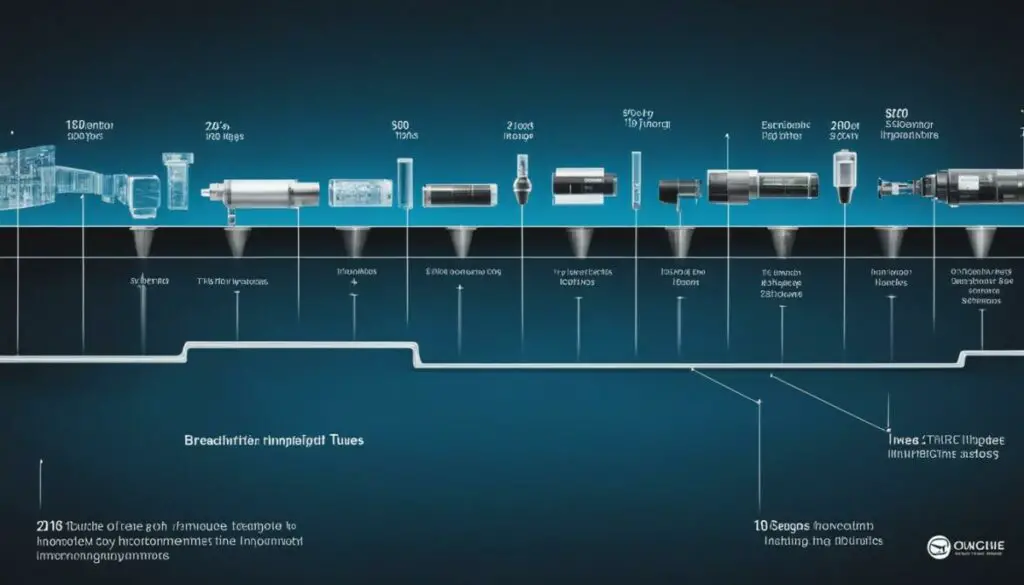
Image intensifier tube technology has evolved significantly since its inception, paving the way for advancements in low-light imaging. Let’s delve into the fascinating development and history of these tubes and discover the breakthroughs that led to their current form.
The Early Years: From Idea to Reality
In the late 1920s, the concept of an image tube was first proposed, sparking interest in the field of low-light imaging. However, it wasn’t until the 1930s that successful infrared converter tubes were created, marking a significant milestone in image intensifier tube development. These early tubes, known as Generation 0 tubes, had limited amplification capabilities but laid the groundwork for future advancements.
World War II and the Rise of Generation 1 Tubes
During World War II, image intensifier tubes took center stage in the military’s night vision applications. Generation 0 image intensifiers were used extensively, but they had one major requirement – an infrared light source. While the technology was revolutionary at the time, further advancements were needed to make these tubes more efficient and adaptable.
The breakthrough came in the 1950s when bialkali antimonide photocathodes were developed. This led to the birth of Generation 1 tubes, which offered significant amplification capabilities. The enhanced sensitivity and quantum efficiency of Generation 1 tubes opened up new possibilities for low-light imaging in various fields.
Generation 2 Tubes and Microchannel Plates
As image intensifier tube technology continued to evolve, Generation 2 tubes entered the scene, introducing a game-changing component – microchannel plates (MCPs). These tiny conductive channels enabled higher levels of amplification, surpassing the capabilities of the previous generations. Generation 2 tubes offered enhanced sensitivity to infrared light, making them even more valuable in military applications and other fields.
Table:
| Generation | Key Advancements |
|---|---|
| Generation 0 | – Infrared converter tubes – Limited amplification capabilities |
| Generation 1 | – Bialkali antimonide photocathodes – Significant amplification capabilities |
| Generation 2 | – Introduction of microchannel plates (MCPs) – Enhanced amplification and sensitivity to infrared light |
The development and history of image intensifier tubes highlight the tireless efforts of researchers and scientists to enhance low-light imaging capabilities. Each generation has brought forth new possibilities and expanded the applications of these remarkable devices. From Generation 0 to Generation 2, image intensifier tubes have undergone continuous development and continue to play a crucial role in various industries and fields where low-light imaging is essential.
Solar Blind Converters for UV Detection
Solar blind converters, also known as solar blind photocathodes, are specialized devices designed to detect ultraviolet (UV) light below 280 nanometers (nm) in wavelength. These converters have the unique ability to detect UV radiation without interference from visible sunlight, making them invaluable for various applications that require sensitivity to UV light.
One of the key components of solar blind converters is the photocathode, which is responsible for the conversion of incoming UV light into detectable signals. An important material used in the production of solar blind photocathodes is cesium telluride. Cesium telluride has exceptional sensitivity to UV light, allowing for accurate and reliable UV radiation detection.
What sets solar blind converters apart from traditional night vision technologies is their specific focus on detecting UV light. While night vision technologies amplify all forms of non-visible light, solar blind converters eliminate interference from the visible spectrum, enabling precise detection and measurement of UV radiation.
Utilizing solar blind converters, researchers and professionals can explore a wide range of applications, including:
- Monitoring and mapping UV radiation levels in the environment
- UV disinfection and sterilization processes
- Industrial and scientific research involving UV-sensitive materials
- UV radiation analysis in medical and dermatological fields
With their unique capability to detect and measure UV light while excluding visible sunlight, solar blind converters play a pivotal role in ensuring accurate UV radiation detection and analysis.
Applications of Solar Blind Converters
| Application | Benefits |
|---|---|
| Environmental Monitoring | Accurate measurement of UV radiation levels in the atmosphere |
| UV Disinfection | Optimizing and validating UV disinfection processes |
| Scientific Research | Analysis of UV-sensitive materials and their behavior |
| Medical and Dermatological Fields | Precise measurement and assessment of UV radiation exposure |
Quote: Exploring the Invisible
“Solar blind converters have revolutionized our ability to detect and analyze ultraviolet light accurately. With their unique design and reliance on cesium telluride photocathodes, these converters enable us to explore the invisible realm of UV radiation while excluding interference from visible sunlight.”
Significant Amplification in Generation 1 Tubes

Generation 1 image intensifiers revolutionized the field of light amplification, thanks to the utilization of multialkali photocathodes. These photocathodes offered increased sensitivity and quantum efficiency in comparison to earlier tube models. The incorporation of thicker layers of the same materials in the S25 photocathode allowed for an extended red response while reducing the blue response, making it particularly suitable for military applications.
The S25 photocathode boasted a sensitivity of approximately 230 μA/lm, enabling the amplification of low-level light sources like moonlight. These advancements marked a significant leap forward in the capabilities of Generation 1 tubes, enhancing both sensitivity and amplification performance.
Advantages of Generation 1 Tubes
Generation 1 image intensifier tubes, equipped with multialkali photocathodes, offered several key advantages:
- Increased sensitivity and quantum efficiency compared to earlier tube models
- Extended red response, enabling enhanced performance in military applications
- Reduced blue response, minimizing undesirable effects in specific scenarios
- Amplification capabilities for low-level light sources, such as moonlight
Comparison: Generation 1 vs. Earlier Image Intensifiers
| Feature | Generation 1 | Earlier Tubes |
|---|---|---|
| Sensitivity | Increased | Limited |
| Quantum Efficiency | Improved | Reduced |
| Red Response | Extended | Standard |
| Blue Response | Reduced | Standard |
| Low-Level Light Amplification | Enabled | Limited |
As illustrated by the table above, Generation 1 tubes represented a significant advancement in terms of sensitivity, quantum efficiency, and red and blue responses. Additionally, they offered the ability to amplify low-level light sources, expanding the scope of potential applications for image intensifier technology. These enhancements provided researchers and practitioners with a powerful tool for various imaging and military purposes.
Micro-Channel Plate Technology in Generation 2 Tubes
In the realm of image intensifier tubes, Generation 2 stands as a significant milestone due to the implementation of microchannel plates (MCP). These tubes leverage the power of MCP in conjunction with the advanced S25 photocathodes, resulting in amplified performance and enhanced sensitivity to infrared light.
Generation 2 tubes offer increased amplification capabilities, making them well-suited for demanding military applications. The integration of MCP, composed of thousands of tiny conductive channels, enables the secondary cascaded emission of electrons, leading to higher levels of amplification. This technology ensures that even minute traces of infrared light can be converted into easily observable visible light.
The addition of microchannel plates to Generation 2 tubes elevates their performance to unprecedented levels. The improved amplification capabilities and enhanced infrared sensitivity of these tubes make them highly sought after in military operations and surveillance activities.
The Role of Microchannel Plates
MCP serves as a vital component in Generation 2 image intensifier tubes, significantly contributing to their increased amplification capabilities. The structure of MCP, comprising an array of micro-scale channels, allows for efficient electron multiplication through secondary electron emission. As each high-energy electron strikes the microchannel plate, a multitude of additional electrons is released through the cascaded emission process, thereby multiplying the original signal.
Generation 2 tubes leverage microchannel plates to achieve higher levels of amplification, ensuring that infrared light is effectively converted into visible light for enhanced imaging capabilities.
The utilization of MCP in tandem with the S25 photocathode further augments the sensitivity of Generation 2 tubes to infrared light. The intensified signal obtained through the microchannel plates facilitates a more comprehensive and detailed interpretation of the infrared spectrum, enabling precise observation and analysis.
With the introduction of microchannel plate technology, Generation 2 image intensifier tubes revolutionized the field of infrared imaging. The efficient amplification capabilities and enhanced sensitivity of these tubes have expanded their applications, particularly in military operations where the ability to detect and convert infrared light is of utmost importance.
Conclusion
The possibility of converting x-ray to infrared opens up exciting opportunities in the field of imaging. Laser-like x-ray beams and chemical processes for converting visible light to infrared energy have shown great promise. These advancements enable us to explore the nanoworld, capture electron motions, and visualize chemical reactions at the atomic level.
Moreover, image intensifier tubes play a crucial role in converting non-visible light sources, such as near-infrared or short-wave infrared, into visible light. This technology has revolutionized low-light processes and facilitated the development of devices like night-vision goggles and medical imaging equipment.
With continuous research and development, we can expect even more innovative applications in the future. From medical imaging and therapeutic treatments to security and surveillance, the x-ray to infrared conversion offers unprecedented possibilities in various fields. The ability to visualize and analyze objects and phenomena with infrared imaging techniques provides valuable insights for scientific study, medical diagnoses, and practical applications.
In conclusion, the conversion from x-ray to infrared is an exciting area of research that will continue to expand the frontiers of imaging technology. With its potential for improved diagnostics, enhanced security measures, and innovative applications, the possibilities are limitless. As scientists and engineers advance their knowledge and refine existing technologies, we can look forward to a future where x-ray to infrared conversion becomes an essential tool in our quest for improved understanding and visualization of the world around us.
FAQ
Can x-rays be converted into infrared?
No, x-rays cannot be directly converted into infrared. X-rays and infrared are two different forms of electromagnetic radiation with distinct properties.
How are laser-like x-ray beams utilized for imaging?
Laser-like x-ray beams have the ability to “see” through materials and capture images at the atomic level. They are generated using ultraviolet lasers and high-harmonic generation, making them ideal for imaging the nanoworld.
Is it possible to convert visible light into infrared energy?
Yes, a chemical process has been developed to convert visible light into infrared energy. This conversion allows for the penetration of living tissue without causing damage, enhancing the reach and effectiveness of therapies such as photodynamic therapy for managing cancer.
How do image intensifier tubes convert light sources?
Image intensifier tubes are optoelectronic devices that can amplify and convert non-visible light sources, such as near-infrared or short-wave infrared, into visible light. They utilize a process of converting light photons into electrons, amplifying these electrons, and then converting them back into photons for visible light output.
How do image intensifier tubes work?
Image intensifier tubes work by utilizing a photocathode to release electrons through the photoelectric effect when light photons strike it. These released electrons are then accelerated and amplified through a microchannel plate before being converted back into photons at a phosphor screen, resulting in visible light that can be viewed.
What is the history of image intensifier tubes?
Image intensifier tubes have evolved over time. They were initially developed in the late 1920s, with the first successful infrared converter tubes created in the 1930s. The introduction of Generation 1 tubes in the 1950s, featuring multialkali photocathodes, marked a significant advancement in sensitivity and amplification. Generation 2 tubes further improved amplification capabilities with the use of microchannel plates.
What are solar blind converters used for?
Solar blind converters are specialized devices designed to detect ultraviolet (UV) light below 280 nanometers (nm) in wavelength. They are particularly useful in applications requiring sensitivity to UV radiation without interference from visible sunlight.
How do Generation 1 image intensifiers differ from earlier tubes?
Generation 1 image intensifiers utilized multialkali photocathodes, providing increased sensitivity and quantum efficiency compared to earlier tubes. The S25 photocathode, with thicker layers of the same materials, extended the red response and reduced the blue response, making it more suitable for military applications.
What is the significance of microchannel plates in Generation 2 tubes?
Generation 2 image intensifiers introduced the use of microchannel plates (MCP) in conjunction with the S25 photocathode. These tubes offered enhanced infrared sensitivity and extended red response, making them even more suitable for military applications. The MCP facilitated the secondary cascaded emission of electrons, resulting in higher levels of amplification.
How can x-ray to infrared conversion impact imaging applications?
The conversion of x-ray to infrared holds the potential to revolutionize various imaging applications. Laser-like x-ray beams, chemical processes to convert visible light to infrared energy, and image intensifier tubes all contribute to expanding the possibilities in imaging and detection techniques, leading to more effective medical treatments and other applications.

